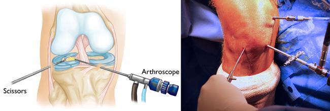
Knee Arthroscopy
Knee arthroscopy is a surgical procedure that allows doctors to view the knee joint without making a large incision (cut) through the skin and other soft tissues. Arthroscopy is used to diagnose and treat a wide range of knee problems.
Your knee is the largest joint in your body and one of the most complex. The bones that make up the knee include the lower end of the femur (thighbone), the upper end of the tibia (shinbone), and the patella (kneecap).
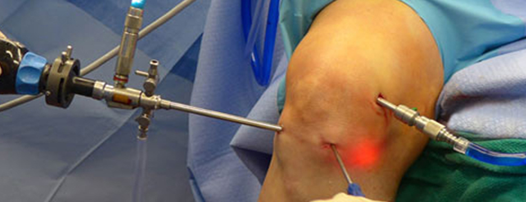
During knee arthroscopy, we inserts a small camera, called an arthroscope, into your knee joint. The camera displays pictures on a video monitor, and we use these images to guide miniature surgical instruments.
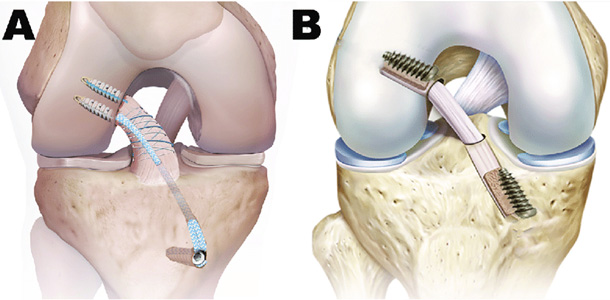
ACL Reconstruction Surgery
ACL surgery is a repair or reconstruction of the anterior cruciate ligament (ACL). The ACL is an important soft-tissue structure in the knee that connects the femur to the tibia. A partially or completely torn ACL is a common injury among athletes.
Partial and complete ACL tears
To determine whether a tear is partial or complete, a doctor will perform two manual tests:
- Lachman test: The physician will try to pull the shin bone away from the thigh bone. If the ACL is torn but still intact, the bones won’t move or will do so only slightly.
- Pivot shift test: The patient lies on their back while the doctor lifts their leg and places rotational pressure on the knee. If the bones don’t shift, the test is negative.
In patients who have only a partial tear, it may be recommended to delay surgery and first see if the ligament heals without it.
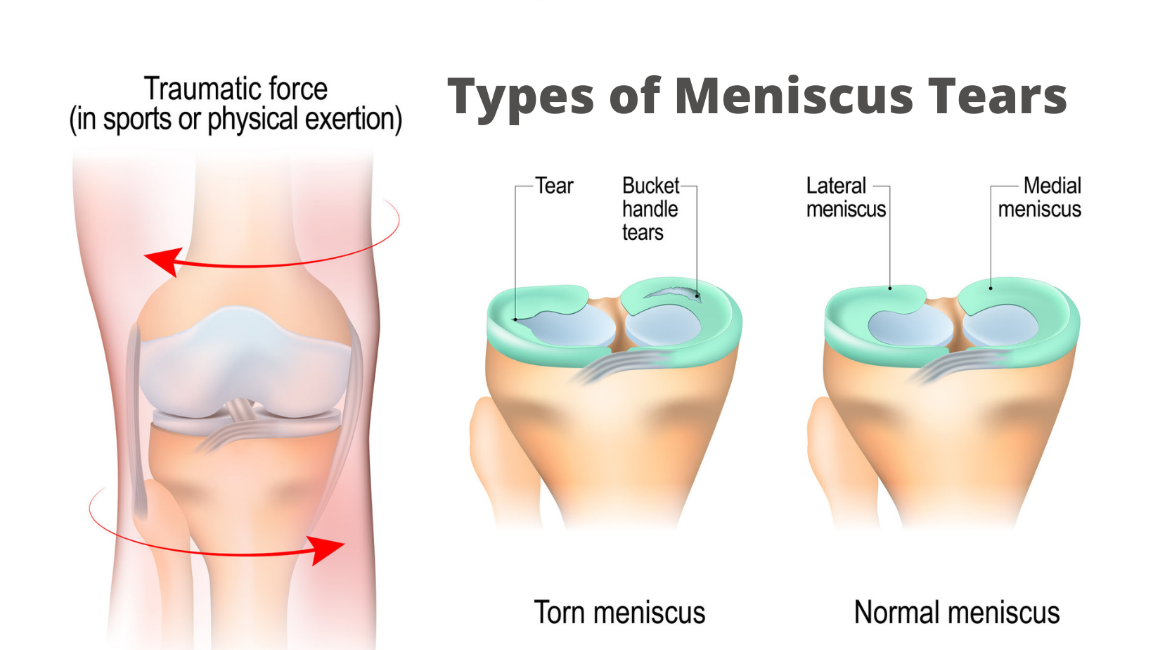
Meniscus Surgery
Meniscus surgery is an operation to remove or repair a torn meniscus, a piece of cartilage in the knee.
Meniscus injury and surgery are common, especially among people who play sports. A sudden twist, turn or collision can tear a meniscus.
Does every meniscus injury need surgery?
Some people need surgery for a torn meniscus, but some don’t. The decision depends on:
- Type, size and location of the tear.
- Your age.
- Your activity level and lifestyle.
- Related injuries (e.g., ACL tear).
- Presence of symptoms (pain, swelling, locking, buckling, etc)

MPFL Reconstruction Surgery
The medial patellofemoral ligament (MPFL) is most commonly injured during a high-impact sporting event that includes pivoting or a tackle. The injury occurs when the patella dislocates and tears the ligament on the inside of the knee and surgery may be required to correct it. Knee MPFL reconstruction surgeon, Dr. Nikhil Verma provides diagnosis along both surgical and nonsurgical treatment options for patients in Chicago who have sustained an MPFL tear. Contact Dr. Susheel's team today!
Although most MPFL tears are caused by a traumatic dislocation, some tears can occur without an obvious injury. Symptoms of an injured or torn MPFL may include:
- Pain in the kneecap, especially when palpated or with activity
- Swelling in the knee after activity
- Feeling of instability or of the knee “giving way”
- Stiffness, locking or pain after sitting for a long time
- Feeling the kneecap slip, especially while turning
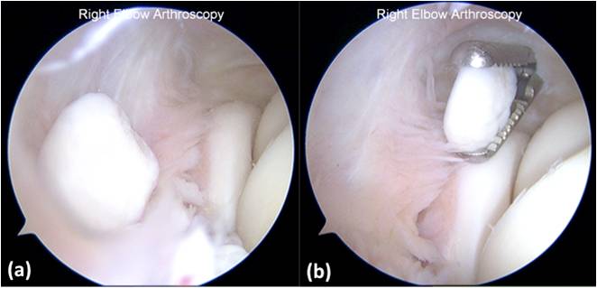
Loose Body Removal
Loose Bodies are fragments of bone and/or cartilage that freely float in the joint space. They may occur singly or in groups. Individuals with a degenerative joint disease (e.g. Arthritis) or traumatic conditions (e.g. Osteochondritis Dissecans) are more likely to develop Loose Bodies in the Knee.
Larger Loose Bodies are typically calcified and thus are easily visible on a plain film X-ray of the affected joint. Loose Bodies that are small or contain little or no bone may not be visible with an X-ray and are typically diagnosed using either CT or Arthrography. MRI may be useful in determining whether associated bone changes have occurred.
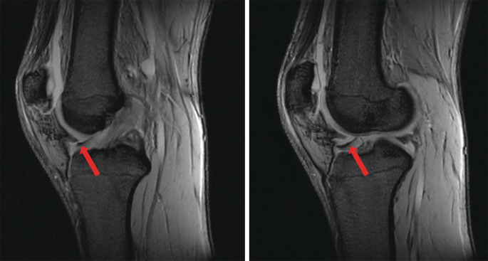
Plica
A plica is a fold in the thin tissue that lines your knee joint. Most people have four of them in each knee. They let you bend and move your leg with ease.
One of the four folds, the medial plica, sometimes gets irritated from an injury or if you overuse your knee. This is known as plica syndrome. It can happen over time to people who run, ride a bike, or use a stair machine, or if you start exercising more than usual. It can also come after trauma to your knee, like bumping it on the dashboard during a car accident.
Synovectomy
Synovectomy is surgery to remove part or all of the synovium, a layer of connective tissue that lines the inside of your joints. The synovium releases fluid and nutrients to keep your joints healthy and moving smoothly.
When you have rheumatoid arthritis (RA) or another type of inflammatory arthritis, your synovium can get swollen and painful. This inflammation is called synovitis. An inflamed synovium makes too much fluid, which wears away the cartilage that cushions your joint. Without cartilage, the bones around the joint rub painfully against each other.
Taking out the inflamed tissue doesn’t cure RA. But it can slow cartilage damage and improve joint pain, at least for a while.
